European Society of Cardiology (ESC) Congress 2016
Optical coherence tomography optimizes PCI results
2016-10-17
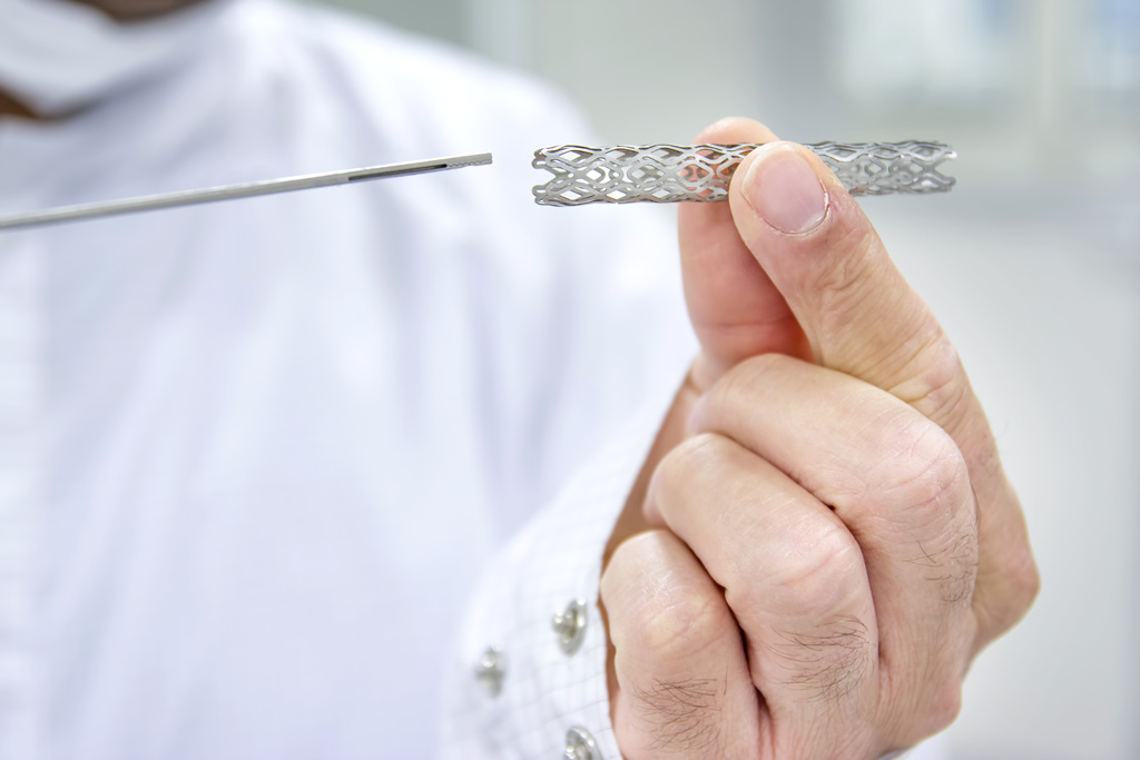
Optical coherence tomography (OCT) has been shown to optimize the results of PCI compared with standard coronary angiography by giving a clearer picture of the coronary arteries during stenting.
This additional information had a direct impact on physician decision-making, leading to procedure optimization in 50 percent of the cases and an improvement in fractional flow reserve (FFR), according to results of the DOCTORS (Does Optical Coherence Tomography Optimise Results of Stenting?) trial presented at the European Society of Cardiology Congress 2016 in Rome, Italy. [Circulation 2016, http://dx.doi.org/10.1161/CIRCULATIONAHA.116.024393]
The study included 240 patients with non–ST-segment elevation acute coronary syndrome (NSTE-ACS) recruited across nine universities in France. Patients were randomized 1:1 to undergo standard angiography-guided PCI or OCT-guided PCI. OCT was performed an average of 3.8 times before, during and after the procedure.
“OCT uses light to capture three-dimensional images and gives a clearer picture of the coronary arteries prior to stent implantation. It allowed clinicians to see significantly more thrombi [69 vs 47 percent; p=0.0004] and calcifications [45.8 vs 9 percent; p<0.0001] compared with standard angiography,” reported lead investigator Professor Nicolas Meneveau of the University Hospital Jean Minjoz in Besançon, France. “As a result, physicians more frequently treated patients with antiplatelet agents in the OCT group compared with the angiography group [53.3 vs 35.8 percent; p=0.007].”
“FFR at the end of procedure, the study's primary endpoint, was significantly improved with OCT compared with angiography [0.94 vs 0.92; p=0.005], as was the proportion of patients with post-PCI FFR >0.90 [82.5 vs 64.2 percent; p=0.0001],” Meneveau continued.
The improvement in FFR was mainly driven by optimization of stent expansion in the OCT group. Compared with standard angiography, OCT was significantly more likely to reveal stent underexpansion (42 vs 10.8 percent; p<0.0001) and edge dissection (37.5 vs 4 percent; p<0.0001). Stent malapposition and tissue protrusion, which were not visible with standard angiography, were detected in 32 and 79 percent of patients in the OCT group, respectively.
“As a result, the procedure was optimized in 50 percent of patients in the OCT group compared with 22.5 percent of patients in the angiography group [p<0.0001],” said Meneveau. “Poststent overinflation was significantly more common with OCT vs angiography [43 vs 12.5 percent; p<0.0001], and the percentage diameter of residual stenosis was lower in the OCT group [7 vs 8.7 percent; p=0.01].”
Although patients in the OCT group were exposed to contrast medium and fluoroscopy for a longer period, the rate of complications such as MI and acute kidney injury did not increase.
“Whilst post-PCI FFR improved with OCT, the trial was conducted in a low-risk NSTE-ACS population with only single lesions. Further studies are required to clarify standardized treatment targets for pre- and post-PCI OCT guidance,” pointed out discussant Professor Stephan Windecker of the Bern University Hospital, Switzerland
| 이전글 | A role for lipoprotein apheresis in refractory angina |
|---|---|
| 다음글 | ICD does not reduce all-cause mortality in non-ischaemic heart failure |


